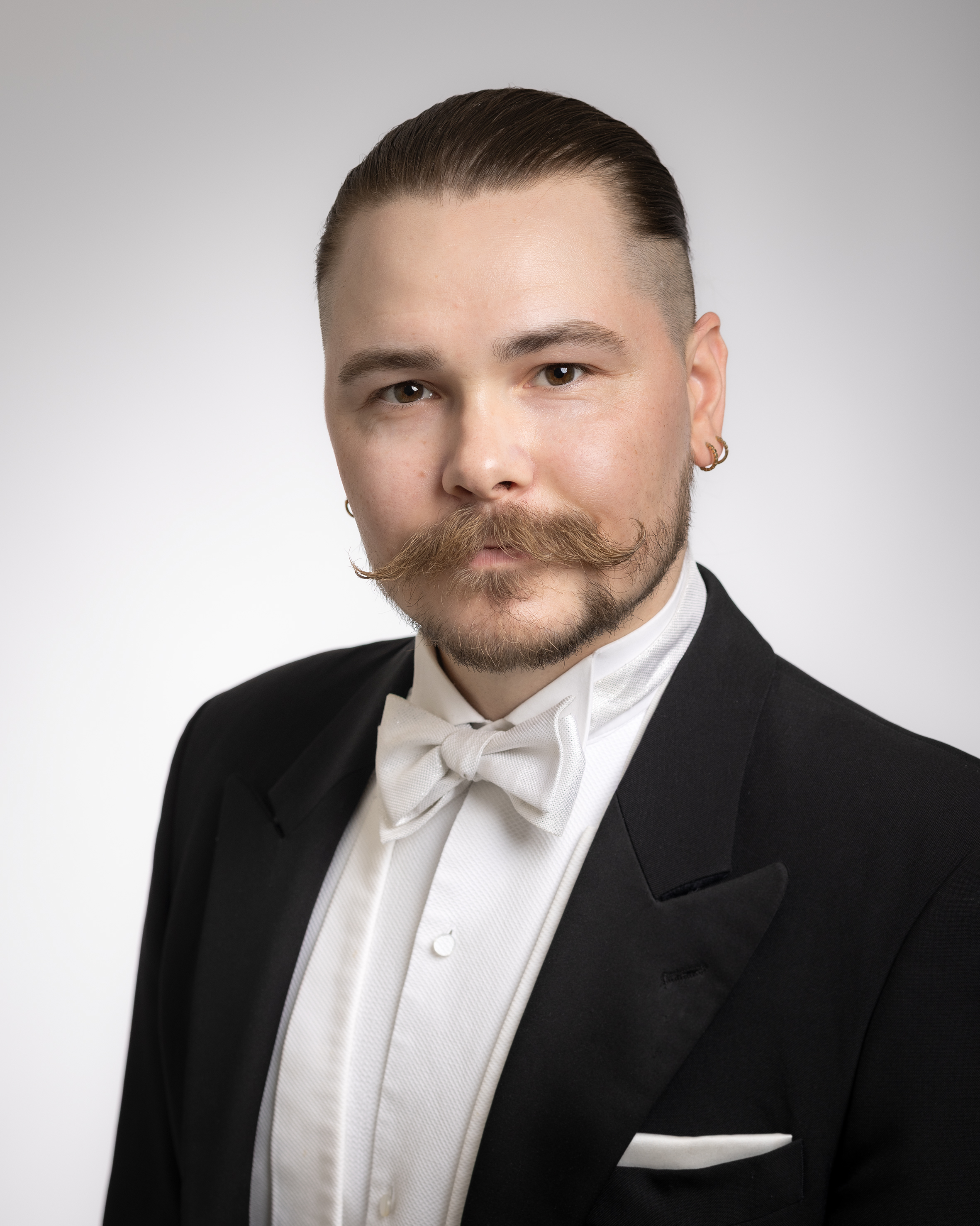
The doctoral dissertation in the field of Medical Physics will be examined at the Faculty of Science, Forestry and Technology, Kuopio campus and online.


What is the topic of your doctoral research? Why is it important to study the topic?
T2 relaxation time mapping is a useful magnetic resonance imaging (MRI) method for assessing articular cartilage health. It is sensitive to the changes typically occurring in cartilage degeneration. Unfortunately, T2 is also highly sensitive to the physical orientation and geometry of the tissue, which can even lead to misdiagnosis if the phenomenon is not considered properly. In the thesis, a method based on splitting T2 into its components using multi-orientation measurements was developed to overcome the orientation-dependency of T2. The T2-derived parameters studied in the thesis are potentially also sensitive to the degenerative changes in cartilage.
Finally, the reproducibility of T2 mapping in a human knee and a standardized phantom was studied using an exceptionally large scanner population of 36 scanners at 16 different hospitals. My knee, which was scanned in this study might even hold the record for most knee MR scans conducted in Finland.
What are the key findings or observations of your doctoral research?
The thesis is split into three studies. The results in the first study demonstrated that precise multi-orientation measurements enable defining new T2-derived orientation-independent parameters. These parameters have great potential to be sensitive to the changes that are typically seen in cartilage degeneration. In the second study, the role of these parameters in cartilage degeneration was studied. Additionally, the parameters were compared with established reference methods for cartilage characterization. Especially the anisotropic, i.e., orientation-dependent component of T2 was found to be sensitive to cartilage degeneration. Additionally, connections to the reference methods were found in the form of various correlations.
In the third study, the reproducibility of T2 was found to be at an acceptable level for knee T2 mapping and at great level in phantom imaging. The unavoidable variation in the scanning parameters and environment necessarily introduces some variation in the measurements. The field strength was not found to affect the T2 reproducibility. On the other hand, also biological variation and day-to-day changes in tissue play their role. In the end, the same kind of variation is also present in everyday situations in the clinical world. Hence, the results describe the real-world situation well.
How can the results of your doctoral research be utilised in practice?
The results related to the T2-derived parameters add to the scientific understanding of the typical changes that are seen in articular cartilage degeneration. They have great potential when methods for early detection of osteoarthritis are being developed.
The reproducibility analysis in the third study answers the question on how reproducible T2 mapping is in the clinical environment when a wide variety of scanners and imaging practices are used. Just like in clinical practice. The population of scanners and imaging units used in the study is a comprehensive overview of scanners and methods used in Finland currently. The results of this final study will be provided for each of the participating hospitals for their use. The imaging units may then use this information when improving the T2 mapping protocols in clinical use.
What are the key research methods and materials used in your doctoral research?
The first two studies were conducted using a high-field-strength (9.4 T) pre-clinical MRI scanner at the AIV-Institute. First, the multi-orientation measuring technique was developed utilizing bovine samples, after which the significance of the defined parameters was assessed in sample group containing human cadaver samples with different levels of cartilage degeneration. The third study was conducted in a close collaboration with Mikkeli central hospital where I worked for the years 2023 and 2024 as a specializing hospital physicist. The study collaborated with almost all of the central and university hospital in Finland. The study is also a part of a larger project focusing on assessing the MR image quality in Finland.
The thesis project was conducted at the Department of Technical Physics at University of Eastern Finland, in the magnetic resonance imaging research group lead by Professor Mikko Nissi.
The doctoral dissertation of Henri Leskinen, MSc, entitled Relaxation time mapping techniques for characterization of articular cartilage and reproducibility analysis of current techniques will be examined at the Faculty of Science, Forestry and Technology, Kuopio campus. The opponent will be Associate Scientist and Associate Professor Matthew Koff, Hospital for Special Surgery, USA, and the custos will be Professor Mikko Nissi, University of Eastern Finland. Language of the public defence is English.
For further information, please contact:
Henri Leskinen, [email protected]